


Spectra of “white LEDs” are characterized by an intense emission in the blue region of the visible spectrum, absent in daylight spectra.
This blue component and the high intensity of emission are the main sources of concern about the health risks of LEDs with respect to their toxicity to the eye and the retina.
The aim of our study was to elucidate the role of blue light from LEDs in retinal damage. Commercially available white LEDs and four different blue LEDs (507, 473, 467, and 449nm) were used for exposure experiments on Wistar rats.
Immunohistochemical stain, transmission electron microscopy, and Western blot were used to exam the retinas. We evaluated LED-induced retinal cell damage by studying oxidative stress, stress response pathways, and the identification of cell death pathways.
LED light caused a state of suffering of the retina with oxidative damage and retinal injury. We observed a loss of photoreceptors and the activation of caspase-independent apoptosis, necroptosis, and necrosis.
A wavelength dependence of the effects was observed. Phototoxicity of LEDs on the retina is characterized by a strong damage of photoreceptors and by the induction of necrosis.
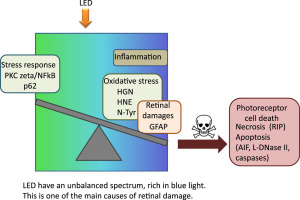
Graphical abstract

Introduction
Retinal degenerations, whether genetic or not, involve death of photoreceptors. Environmental factors such as smoking, obesity, and hypertension are important in the progression of the different retinopathies [1].
In addition, the exposure to intense natural or artificial light can be detrimental to eye tissues by causing photochemical damages. Many studies have been conducted to evaluate the effect of light on the evolution of preexisting retinopathy [2].
For the last few years this interest has been concentrated on age-related macular degeneration (AMD), a leading pathology of the elderly in Western countries [3].
After many conflicting studies it was concluded that light is a risk factor in the early stages of AMD, although it is not recognized as an aggravating factor for the pathology [4], [5], [6].
The retinal damages induced by light depend on radiation intensity, radiation wavelength, and time of exposure [7], [8]. Light-induced retinal lesions are characterized by a degeneration of photoreceptors outer segments leading to their death by apoptosis [9], [10].
Recently, the use of new technologies in domestic lighting induced a renewal of the interest on the effects of light on the retina.
CLICK HERE TO READ MORE FROM THE REPUBLICAN VOICE
Among these new devices, light emitting diodes (LEDs) are at the center of this interest. From a technical point of view LEDs have many advantages such as low energy consumption, high mechanical strength, and, especially, long life.
One of the major concerns with the use of this technology is the emission spectrum of LEDs. The spectrum of LEDs is characterized by an intense blue light component absent in the daylight spectra [11].
In recent years, governmental agencies such as the ANSES (French Agency for Food, Environmental and Occupational Health and Safety) in 2010 stressed the need for extensive experimental studies on the phototoxicity of LEDs to confirm or refute the fears raised by researchers, ophthalmologists, and physicists concerning the intensive use of these devices.
Actually, the most commonly used LEDs present a high luminance level and generate a visual discomfort related to the “punctual” character of the emitting surfaces.
In addition, the spectral imbalance of white LEDs available on the market, due to the production of white light by the processing of a high-frequency radiation and reemission in a complementary wavelength, exposes the eye to residual short wavelength radiations, the most dangerous to the retina.
This problem could be increased by a reduction of the pupillary contraction, stimulated at around 480 nm, a region of low emission in the spectrum of some LEDs, resulting in an increase of retinal light exposure.
In this study, we investigate, in rats, the molecular mechanism of retinal degeneration by using five different LEDs: white LEDs, green LEDs (507 nm), and three blue LEDs (449, 467, 473 nm) to evaluate the role of blue radiation in cellular damage and the progression of retinal lesions.
Section snippets
Animals
Six-week-old male Wistar rats were purchased from Janvier labs and kept 1 week for adaptation in our animal facilities in a light cycle of 12 h. All procedures were in compliance with the animal use and care committee of the National Veterinary School of Alfort.
Light source
We used two types of lighting devices built and characterized by the Lighting and Electromagnetism Division of the Scientific and Technical Center for Building (CSTB, Saint Martin d׳Heres, France): the first contains only white LEDs
Dilated fundus examination (DFE) showed no bleaching of the retina
DFE is a diagnostic procedure used to evaluate the internal ocular health. Until today, regulations of light toxicity on the retina have established ELV (exposure limit values) [14].
These ELV are based on a study of fundus examination following a single exposure to light for 8 h. They establish that light is toxic to the eye when there is a bleaching of the retina.
In our study, DFE was performed just after exposure by a veterinary ophthalmologist and two assistants. No bleaching of the retina
Discussion
In this paper we analyzed the effects of the so-called “white LEDs” and blue-enriched LEDs on the retina. We show an important damage of the photoreceptors layer after 18 h of exposure.
More importantly, the analysis, at the molecular level, of the effectors of cell death shows an activation of necrotic cell death, a rare event in this type of degeneration.
The current regulation [26] establishes that light is toxic to the eye when there is a bleaching of the retina as seen by fundoscopy. In our
Acknowledgments
CLICK HERE TO READ MORE FROM THE REPUBLICAN VOICE
This work was supported by INSERM, ENVA, CSTB, and by an ADEME financial support (RETINALED project). I.J. has a fellowship from ADEME.
The authors are indebted to Dr. Patricia Lassiaz for helpful discussions on PKC zeta and to Mrs. Brigitte Goldenberg and Mr. Christophe Klein for technical assistance.